The Joan Mecsas Lab
Yersinia - Host Interaction
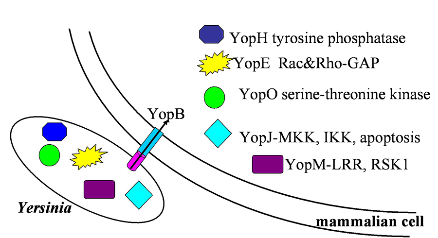
My laboratory studies the bacterial pathogen, Yersinia, which is virulent in a variety of mammals, including mice and humans. All pathogenic Yersinia species, including Y. pseudotuberculosis, Y. enterocolitica and Y. pestis, encode a type III secretion system (TTSS) which injects bacterial effector proteins, called Yops, from Yersinia into mammalian cells. In cultured cells, the 5 effector Yops disable or alter normal signal-transduction pathways once they are injected into the cells. For example, YopE has GAP activity on RhoA and Rac1 and prevents phagocytosis of Yersinia . In fact, several of the Yops have been shown to bind to and inactivate several mammalian protein targets.
Figure 1. This diagram illustrates the function of the five Yersina Yop proteins. As described above, the bacterium introduces these molecules into cells to facilitate its growth.
Yop Signaling Mechanisms and Host Defense
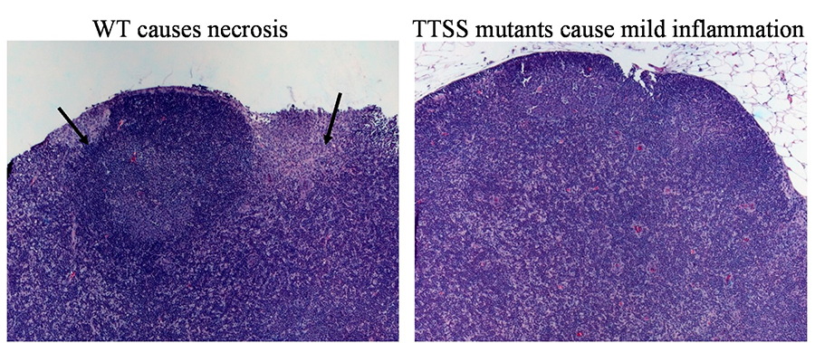
Our work focuses on identifying the immune cell targets of and the signal-transduction pathways that are inactivated by Yops during infection. We have used a variety of mouse model systems of infection, including oral, intranasal and intravenous infections, to identify which Yops are required for Yersinia to colonize and replicate in various host tissues. Furthermore, we have investigated the behavior of yop mutants in mice defective in specific aspects of immunity and the host immune response to infection with wildtype and yop mutants. We have found that different Yops are important in different tissues and that Yops target different host defenses. We are currently investigating the signal transduction mechanisms targeted by the Yops in these various cells in infected tissues.
Figure 2. Yops affect host - Yersinia interaction in many ways. The inflammatory response is one of these effects. One way this can occur is illustrated by the altered response to TTSS mutants.
Yop Translocation Mechanisms
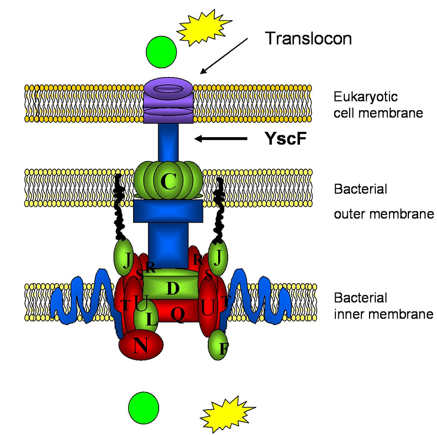
A second major project focuses on understanding the process of translocation of Yops into mammalian cells. Using a combination of genetics, biochemistry and high throughput screens, we are dissecting the interface between the needle and the translocon and investigating the molecular events that occur for productive translocation of Yops into mammalian cells.
Figure 3. This illustration depicts the components of the Type III Secretion system. This system translocates the Yops from Yersinia into the host cell.


