Astrocytes, Aging, and Neural Stability: An MBR Thesis on Brain Homeostasis
As part of a Tufts GSBS research spotlight series, recent MBR graduates discuss their thesis projects, key findings, and the skills they developed through independent, PhD‑level research training.

By Ellen Sojka, MBR26
Member of: The Chris Dulla Lab
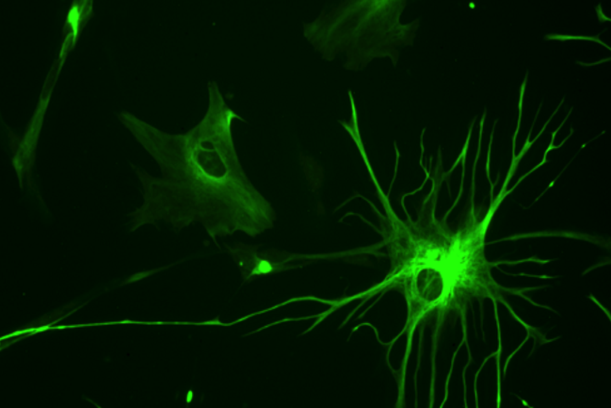
I have always been fascinated by how the brain maintains stability in the face of constant activity, aging, and injury—and how this balance breaks down in disease. That curiosity led me to my master’s thesis project studying astrocytes, a type of glial cell that can be described as the brain’s “homeostatic guardians.” Astrocytes regulate neurotransmission, support the blood–brain barrier, shape inflammatory responses, and help maintain the brain’s chemical environment. One of the most remarkable features of astrocytes is their ability to form large, interconnected networks through structures called gap junctions. These gap junctions allow astrocytes to communicate and share molecules across the brain, helping maintain neural homeostasis.
Astrocytes, Aging, and Atypical Cell States
Because astrocytes are critical regulators of brain function, they have become increasingly implicated in age-related cognitive decline and neurodegenerative disease. While astrocytic protein expression undergoes characteristic, spatially broad changes during normal aging, preliminary data from Dr. Chris Dulla’s lab also established an increasing proportion of astrocytes that sporadically lose expression of key functional proteins, impairing their essential functions. Referred to as atypical astrocytes (AtAs), these cells exhibit dramatic reductions in proteins responsible for clearing glutamate from synapses, buffering potassium, and removing amyloid-beta from the brain. Since these functions are essential for healthy neural signaling, the loss of these proteins may contribute to dysfunction at the network level.
The Research Question
Importantly, AtAs also show decreased levels of connexin 30 and connexin 43, two proteins that form astrocytic gap junctions. This observation raised an important question: if AtAs lose these proteins, do they also lose their ability to functionally connect with neighboring astrocytes?
To investigate this question, I studied whether AtAs become functionally uncoupled from the broader astrocytic network during aging. This work was part of a larger project spearheaded by Panorea Tirja, a fourth-year PhD candidate in the Dulla lab. As one of the few researchers studying AtAs, Panorea provided invaluable mentorship and helped introduce me to this entirely new area of neuroscience research. Under her guidance, I independently led the investigation into astrocytic gap junction coupling in aging brains.
Skills Gained Through Independent Research
Through this project, I gained extensive experience with immunohistochemistry and acute slice electrophysiology. Over the course of my thesis, I learned how to perform whole-cell patch clamp recordings from neurons and astrocytes in aged mouse brain tissue, optimized a staining protocol for thick brain slices, and developed an image analysis pipeline to quantify biocytin diffusion between astrocytes. Because many of these techniques were new to the lab, developing and troubleshooting protocols independently was both challenging and rewarding.
Beyond technical skills, this experience transformed how I think as a scientist. Working on a highly novel project forced me to become more adaptable, rigorous, and creative in my experimental approach. I learned how to critically analyze unexpected results, redesign experiments when necessary, and ask sharper scientific questions. Most importantly, I developed confidence in my ability to lead independent research.
Key Findings and Scientific Impact
My findings revealed that AtAs exhibit approximately 70% lower connexin 30 and connexin 43 expression than typical astrocytes at both young and adult time points, suggesting that AtAs represent a distinct astrocyte state rather than a progressive one. To determine whether this reduction in gap junction protein expression impaired functional coupling, I used a biocytin diffusion assay in acute cortical brain slices followed by post hoc immunolabeling. I found that biocytin diffusion into and out of AtAs was significantly reduced compared to typical astrocytes, indicating impaired coupling of these cells with the astroglial network. AtAs also displayed increased membrane resistance, consistent with broader reductions in ion channel expression.
Together, these findings demonstrate that the loss of connexin proteins in AtAs leads to functional uncoupling from the astrocytic network. This uncoupling may contribute to disruptions in neural homeostasis and communication in the aging brain. More broadly, our work provides new insight into how astrocyte dysfunction may shape both normal aging and neurological disease.
Working at the Edge of Discovery
One of the most exciting aspects of this project was studying something so new. Because only a handful of researchers are currently investigating AtAs, there is very little existing literature to guide the work. While that uncertainty could sometimes feel intimidating, it ultimately deepened my passion for research. Every experiment raised new questions: Are AtAs present in other neurological disorders, such as epilepsy? Do AtAs represent a temporary or more permanent state in aging? How do increasing numbers of AtAs affect neural circuits and cognition over time?
Working in such an unexplored field reminded me just how much remains unknown in neuroscience, and how meaningful even small discoveries can be for understanding human health and disease.
How the MBR Program Supported My Growth
The MBR program provided the support and training necessary for me to thrive throughout this research experience. Opportunities to rotate through multiple labs, present at journal clubs, and develop specific aims for my thesis helped build both my scientific foundation and my confidence as a researcher. Because the program is highly interdisciplinary, discussions with students and faculty across different research areas continually challenged me to think more broadly and critically about science.
I also found myself regularly returning to concepts from coursework and applying them directly to my experiments, which reinforced the strong connection between classroom learning and hands-on research. In addition, the mentorship and support within the MBR program were instrumental in helping me prepare for my next steps. Program director Dr. Peter Juo consistently provided thoughtful guidance throughout the PhD application process, helping students navigate an especially competitive application cycle.
Looking Ahead
This fall, I will begin pursuing a PhD in neuroscience in the Boston University Graduate Program for Neuroscience (GPN). My MBR experience confirmed not only that I am capable of pursuing a research career, but also that I genuinely love the process of scientific discovery. The MBR program gave me the opportunity to immerse myself in meaningful research, and it ultimately helped shape the scientist I hope to become.
Related Links:
MBR Admissions
Request Information
Student Stories
Department:
MS in Biomedical Research