The Philip Haydon Lab
Roles of Astrocytes in Brain Function in Health and Disease
The brain is comprised of a combination of neurons and glia. For much of the past century the focus of studies on brain function examined the important roles neurons play in information processing. In comparison to neurons, glia are electrically silent and their role in the nervous system has been a mystery. In the 1990’s technically advances in imaging and molecular genetics allowed glia to be examined and, as a consequence, we now know that they play pivotal roles in the control of brain and behavior in health and disease.
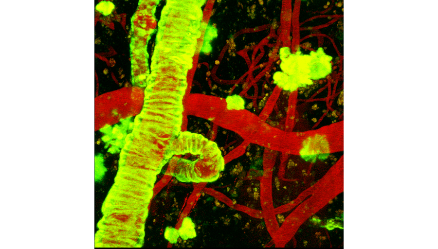
Figure 1. Imaging of Amyloid plaques in vivo in mouse models of Alzheimer’s disease. Images were acquired using two photon microscopy, a technique used in the laboratory to investigate the role of glial cells in brain function in mouse models of depression, epilepsy and Alzheimer’s disease.
Work in Haydon’s laboratory integrates a variety of technical approaches to illuminate roles played by glia. These approaches include molecular genetics, electrophysiology, imaging as well as behavioral studies. In the 1990’s Haydon’s laboratory was the first to discover that astrocytes, a sub-type of glial cell, exhibit the regulated release of chemical transmitters that were previously only thought to be released from neurons. Since that time the focus of the laboratory’s investigations have been on the identification of the consequences of gliotransmission for circuit function and behavior.
In 2009 Haydon’s group applied molecular genetics to the study of the astrocyte and demonstrated that a process termed sleep homeostasis is modulated by the astrocyte. These glial cells regulate extracellular adenosine, a compound that is known to modulate sleep. Through this glial pathway they discovered that the drive to sleep is dependent on this glial signal and that the ability of sleep deprivation to induce sleep and cognitive consequences acts through the astrocyte.
Current studies continue to study how astrocytes regulate synaptic transmission at the tripartite synapse and because sleep is co-morbid with may psychiatric conditions, considerable effort is being placed on studying potential roles of astrocytes in the regulation of mental health.
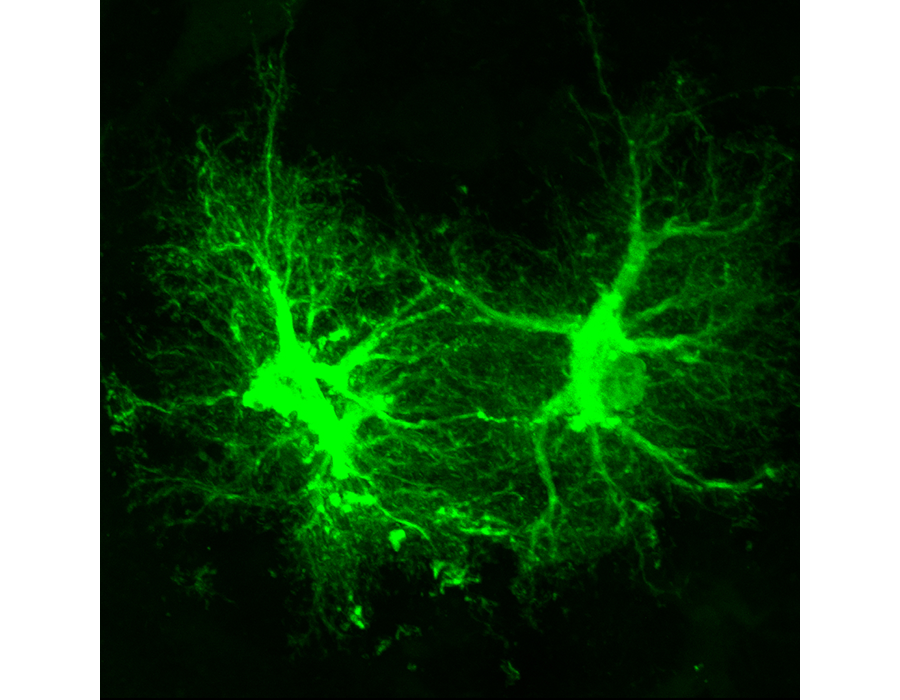
Figure 2. Viral transduction of astrocytes is used to perturb the function of these glial cells to identify their roles in brain function.
Numerous disorders of brain function, including Alzheimer’s disease and Epilepsy, are associated with changes in glial structure and protein expression. However, this is a classic experimental problem – are the changes in glial structure/function contributing to the disorder, or are the changes in glia a consequence of the pathology already present in the brain. Using viral transduction mechanisms Haydon is investigating whether these “reactive” astrocytes lead to altered neuronal function. They have found that alterations in the astrocyte lead to compromised synaptic inhibition which leads to increased excitability. Because these changes in astrocytes are known to occur during the development of epilepsy this raises the exciting potential for a new therapeutic target the treatment of seizure disorders.


