The Pablo Argüeso Lab
Our research is in the fields of glycobiology and ophthalmology, focusing on the structure and function of the glycocalyx—a carbohydrate-rich layer lining the surface of eukaryotic cells.
Fig 1. The ocular surface epithelial glycocalyx. Transmembrane mucins emanating up to 500 nm above the plasma membrane interact with multimeric galectin-3 to promote epithelial integrity. Major classes of glycans at the ocular surface include O-glycans, commonly attached to serine (Ser) or threonine (Thr) amino acids, and N-glycans, attached to asparagine (Asn). Mucins, glycoproteins and glycolipids are embedded in the plasma membrane (Argüeso P, Guzman-Aranguez A, Mantelli F, Cao Z, Ricciuto J, Panjwani N. 2009. J Biol Chem. 28, 23037-23045; Taniguchi T, Woodward AM, Magnelli P, et al. 2017. J Biol Chem. 292, 11079-11090; Argüeso P. 2020. Eye Cont Lens. 46:S53-S56).
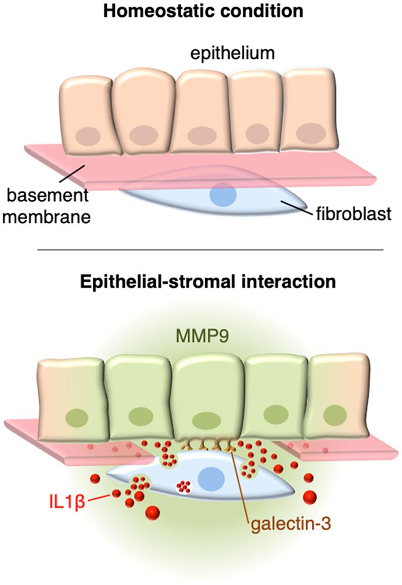
My laboratory has developed innovative methods clearly demonstrating how glycans regulate many aspects of the inflammatory response. These include elucidating the role of endomucin as an anti-adhesive molecule that prevents the rolling of leukocytes on vascular endothelial cells, and how galectins, a group of galactose-binding proteins, promote a proinflammatory environment following ocular trauma.
Fig. 2. Disruption of the basement membrane by trauma allows for direct interactions between epithelial cells and stromal fibroblasts, resulting in a cascade of signaling events critical for tissue repair. Galectin-3 of epithelial origin initiates such events by interacting with fibroblasts. Consequently, the stromal fibroblasts synthesize and secrete IL-1β, which act in a paracrine fashion to regulate the proteolytic microenvironment by activating the MMP9 promoter in epithelial cells. This reciprocal form of communication leads to the remodeling of the extracellular matrix (AbuSamra DB, Mauris J, Argüeso P. 2019. Sci Signal. 12(590):eaaw7095. doi: 10.1126/scisignal.aaw7095.).


